Human Periostin ELISA | BI-20433
-
Method
Sandwich ELISA, HRP/TMB, 12×8-well detachable strips
-
Sample type
Serum, plasma (EDTA, citrate, heparın), urine, cell culture supernatant
-
Sample volume
10 µl / well
-
Assay time
2 h / 2 h / 1 h / 30 min
-
Sensitivity
20 pmol/l (= 1 800 pg/ml)
-
Standard range
0 – 4 000 pmol/l (= 0 – 360 000 pg/ml)
-
Conversion factor
1 pmol/l = 91 pg/ml (MW: 91 kDa)
-
Specificity
Endogenous and recombinant human Periostin; all known isofoms.
-
Precision
In-between-run (n=10): ≤ 6 % CV
Within-run (n=5): ≤ 3 % CV
-
Cross-reactivity
Due to the high sequence homology between human Periostin and Periostin of other species there is potential cross-reactivity with cynomolgous monkey, dog and cat Periostin.
-
Use
Research use only
-
Validation Data
See validation data tab for: precision, accuracy, dilution linearity, values for healthy donors, etc
Human Periostin ELISA Product Overview
The human Periostin ELISA kit is a 5.5 hour, 96-well sandwich ELISA for the quantitative determination of Periostin in human serum and plasma. The Periostin assay employs human serum-based standards to ensure the measurement of biologically reliable data.
The human Periostin kit uses highly purified, epitope mapped antibodies with characterized binding kinetics.
Human Periostin ELISA Assay Principle
The Periostin ELISA kit human is a sandwich enzyme immunoassay for the quantitative determination of periostin in human serum and plasma samples.
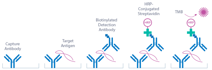
The figure below explains the principle of the Periostin ELISA:
Capture antibody: monoclonal mouse anti-human Periostin antibody
Detection antibody: polyclonal goat anti-human Periostin antibody, biotin labeled
Target antigen: human Periostin
In a first step, pre-diluted standard/control/sample and biotinylated polyclonal goat anti-human Periostin antibody are pipetted into the wells of the microtiter strips, which are pre-coated with mouse monoclonal anti-human Periostin antibody. Periostin present in the standard/control/sample binds to the pre-coated antibody in the well and forms a sandwich with the detection antibody. In a washing step all non-specific unbound material is removed. In a second step, the conjugate (streptavidin-HRP) is pipetted into the wells and reacts with the detection antibody. After another washing step, the substrate (TMB, tetramethylbenzidine) is pipetted into the wells. The enzyme-catalyzed color change of the substrate is directly proportional to the amount of Periostin present in the sample. This color change is detectable with a standard microtiter plate reader. A dose response curve of the absorbance (optical density, OD at 450 nm) vs. standard concentration is generated, using the values obtained from the standard. The concentration of human Periostin in the sample is determined directly from the dose response curve.
Human Periostin ELISA Typical Standard Curve
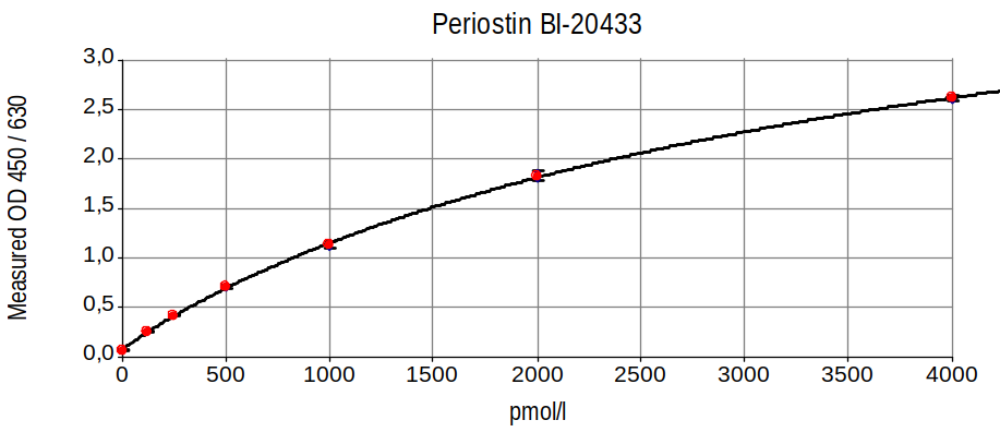
The figure below shows a typical standard curve for the human Periostin ELISA. The immunoassay is calibrated against recombinant human Periostin peptide:
Human Periostin ELISA Kit Components
| Contents | Description | Quantity |
|
PLATE |
Monoclonal mouse anti-human Periostin antibody pre-coated microtiter strips in a strip holder, packed in an aluminum bag with desiccant | 12 x 8 tests |
| WASHBUF |
Wash buffer concentrate 20x, natural cap |
1 x 50 ml |
| STD | Standards 1-7, (0, 125, 250, 500, 1000, 2000, 4000 pmol/l), recombinant human Periostin in human serum, white caps, lyophilized | 7 vials |
| CTRL | Control A and B, yellow cap, lyophilized, exact concentrations see labels | 2 vials |
| ASYBUF | Assay buffer, red cap, ready to use | 1 x 55 ml |
| AB | Polyclonal goat anti-human Periostin antibody – biotin labeled, green cap, ready to use | 1 x 18 ml |
| CONJ |
Conjugate (streptavidin-HRP), amber bottle, amber cap, ready to use |
1 x 18 ml |
| SUB |
Substrate (TMB solution), amber bottle, blue cap, ready to use |
1 x 22 ml |
| STOP |
Stop solution, white cap, ready to use |
1 x 7 ml |
Storage instructions: All reagents of the human Periostin ELISA kit are stable at 4°C (2-8°C) until the expiry date stated on the label of each reagent.
Sample Collection & Storage
Serum, EDTA plasma, heparın plasma, citrate plasma, urine and cell culture supernatant are suitable for use in this Periostin assay. Do not change sample type during studies. We recommend duplicate measurements for all samples, standards and controls. The sample collection and storage conditions listed are intended as general guidelines.
Serum & Plasma
Collect venous blood samples in standardized serum separator tubes (SST) or standardized blood collection tubes using EDTA, heparın or citrate as an anticoagulant. For serum samples, allow samples to clot for 30 minutes at room temperature. Perform separation by centrifugation according to the tube manufacturer’s instructions for use. Assay the acquired samples immediately or aliquot and store at -25°C or lower. Lipemic or haemolyzed samples may give erroneous results. Samples are stable for four freeze-thaw cycles. Thawed samples should be assayed as soon as possible.
Urine
Note: the experiments performed to measure Periostin in urine samples did not undergo a full validation according to FDA/ICH/EMEA guidelines. However, our performance check suggests that urine samples can be measured with this ELISA.
Aseptically collect the first urine of the day (mid-stream), voided directly into a sterile container. Centrifuge to remove particulate matter, assay immediately or aliquot and store at -25°C or lower. Do not freeze-thaw samples more than four times. Thawed samples should be assayed as soon as possible.
Cell Culture Supernatant
Note: the experiments performed to measure Periostin cell culture supernatant samples did not undergo a full validation according to FDA/ICH/EMEA guidelines. However, our performance check suggests that cell culture supernatant samples can be measured with this ELISA.
Remove particulates by centrifugation and assay immediately or aliquot and store samples at
-25°C or lower. Do not freeze-thaw samples more than four times. Thawed samples should be assayed as soon as possible.
Reagent Preparation
Wash Buffer
| 1. | Bring the WASHBUF concentrate to room temperature (18-26°C). Crystals in the buffer concentrate will dissolve at room temperature. |
| 2. | Dilute the WASHBUF concentrate 1:20, e.g. 50 ml WASHBUF + 950 ml distilled or deionized water. Only use diluted WASHBUF when performing the assay. |
The diluted WASHBUF is stable up to one month at 4°C (2-8°C).
Standards & Controls
| 1. | Pipette 200 µl of distilled or deionized water into each standard (STD) and control (CTRL)vial. The exact concentration is printed on the label of each vial. |
| 2. | Leave at room temperature (18-26°C) for 20 min. Vortex gently. |
Reconstituted STDs and CTRLs are stable at -25°C or lower until expiry date stated on the label. STDs and CTRLs can undergo one freeze-thaw cycle.
STDs/CTRLs must be diluted 1+50 with ASYBUF (assay buffer) prior to the assay, e.g. 10 µl STD/CTRL + 500 µl ASYBUF.
Note: 150 µl pre-diluted STD/CTRl is needed per well.
Sample Preparation
Bring samples to room temperature and mix samples gently to ensure the samples are homogenous. We recommend duplicate measurements for all samples.
Serum and plasma samples must be diluted 1+50 with ASYBUF (assay buffer) prior to the assay, e.g. 10 µl sample + 500 µl ASYBUF. 1+50 diluted serum and plasma samples for which the OD still exceeds the highest point of the standard range can be further diluted with ASYBUF.
Note: 150 µl pre-diluted sample is required per well.
Use urine and cell culture supernatant samples undiluted. Urine and cell culture supernatant samples for which the OD exceeds the highest point of the standard range can be diluted 1+50 with ASYBUF (assay buffer).
Human Periostin ELISA Assay Protocol
Read the entire protocol before beginning the assay.
| 1. | Bring samples and reagents to room temperature (18-26°C). |
| 2. | Mark positions for STD/CTRL/SAMPLE (standard/control/sample) on the protocol sheet. |
| 3. | Take microtiter strips out of the aluminum bag. Store unused strips with desiccant at 4°C in the aluminum bag. Strips are stable until expiry date stated on the label. |
| 4. | Add 150 µl pre-diluted (1+50) STD/CTRL/SAMPLE into the respective wells, swirl gently.
Note: use urine and cell culture supernatant samples undiluted. |
|
5. |
Cover the plate tightly and incubate for 2 hours at room temperature (18-26°C). |
| 6. | Aspirate and wash wells 5 x with 300 µl diluted WASHBUF (wash buffer). After the final wash, remove the remaining WASHBUF by strongly tapping plate against a paper towel. |
| 7. | Add 150 µl AB (biotinylated anti-human Periostin antibody, green cap) into each well, swirl gently. |
| 8. | Cover tightly and incubate for 2 hours at room temperature. |
| 9. | Aspirate and wash wells 5 x with 300 µl diluted WASHBUF. After the final wash, remove the remaining WASHBUF by strongly tapping plate against a paper towel. |
| 10. | Add 150 µl CONJ (conjugate, amber cap) into each well. Swirl gently. |
| 11. | Cover tightly and incubate for 1 hour at room temperature. |
| 12. | Aspirate and wash wells 5 x with 300 µl diluted WASHBUF. After the final wash, remove the remaining WASHBUF by strongly tapping plate against a paper towel. |
| 13. | Add 150 µl SUB (substrate, blue cap) into each well. |
| 14. | Incubate for 30 min at room temperature in the dark. |
| 15. | Add 50 µl STOP (stop solution, white cap) into each well. Swirl gently. |
| 16. | Measure absorbance immediately at 450 nm with reference 630 nm, if available. |
Calculation of Results
Construct a standard curve from the absorbance read-outs of the standards using commercially available software capable of generating a four-parameter logistic (4-PL) fit. Alternatively, plot the standards’ concentration on the x-axis against the mean absorbance for each standard on the y-axis and draw a best fit curve through the points on the graph. Curve fitting algorithms other than 4-PL have not been validated and will need to be evaluated by the user.
Obtain sample concentrations from the standard curve. If required, pmol/l can be converted into pg/ml by applying a conversion factor (1 pg/ml = 0.011 pmol/l (MW: 91 kDa)). Sample dilutions above 1+50 have to be considered when calculating the final concentration of the sample. For undiluted samples, divide values by 50 to obtain the final sample concentration.
The quality control (QC) protocol supplied with the kit shows the results of the final release QC for each kit lot at production date. Data for OD obtained by customers may differ due to various influences including the normal decrease of signal intensity throughout shelf life. However, this does not affect validity of results as long as an OD of 1.50 or higher is obtained for the standard with the highest concentration and the values of the CTRLs are within the target range (see labels).
Periostin Protein
Periostin (OSF-2) is secreted as a 91 kDa homodimeric soluble extracellular matrix protein expressed in collagen-rich fibrous connective tissues. There are at least 7 isoforms of Periostin, caused by alternative splicing (http://www.uniprot.org/uniprot/Q15063).
|
Molecular weight |
91 kDa |
| Cellular localisation | Extracellular or secreted |
| Post-translational modifications | Glycosylation, disulphide bonds |
| Sequence similarities | FAS1 superfamily |
| Alternative names | POSTN, OSF-2, OSF2, PDLPN, PN |
| Pubchem ID | 187888 |
| Genecards | POSTN |
| OMIM | 608777 |
| PDB | 5WT7 5YJG 5YJH |
| Pfam | PF02469 |
| Protein Atlas | POSTN |
| Uniprot ID | Q15063 |
Periostin Function
Periostin is involved in osteoblast recruitment, attachment and spreading. It has been associated with the epithelial-mesenchymal transition in cancer and with the differentiation of mesenchyme in the developing heart. Periostin has functions in osteology, tissue repair, oncology, cardiovascular and respiratory diseases, and in various inflammatory settings.
-
Nephrology
Kidney fibrosis (An et al., 2018; Hwang et al., 2017; Mael-Ainin et al., 2014; Sen et al., 2011)
Polycystsic kidney disease (Raman et al., 2018; Wallace et al., 2014)
Hypertensive nephropathy (Guerrot et al., 2012)
Kidney injury (Satirapoj et al., 2015, 2014, 2012)
IgA Nephrophathy (Hwang et al., 2016; Satirapoj et al., 2014)
-
Oncology
Multiple myeloma (Terpos et al., 2016)
Ameloblastoma (Kang et al., 2018)
Ovarian cancer (Ryner et al., 2015; Sung et al., 2016; Tang et al., 2018)
Papillary thyroid carcinoma (Giusca et al., 2017)
Head and neck cancer (Liu et al., 2018)
Breast carcinoma (Kim et al., 2017; Lambert et al., 2016; Nuzzo et al., 2016; Ratajczak-Wielgomas et al., 2017)
Lung cancer (Che et al., 2017; Zhang et al., 2017)
Osteosarcoma (Hu et al., 2016)
Gastric cancer (Liu et al., 2016)
Bladder cancer (Silvers et al., 2016)
Prostate cancer (Tian et al., 2015)
-
Other
Wound healing
Miscarriage (Freis et al., 2017)
Skin diseases (Murota et al., 2017; Sung et al., 2017)
Liver stiffness (Honsawek et al., 2015)
-
Respitatory disease
Asthma (Carpagnano et al., 2018; Górska et al., 2016; Hoshino et al., 2016; Inoue et al., 2016; Kim et al., 2014; Lee et al., 2018; Matsusaka et al., 2015; O’Dwyer and Moore, 2017; Song et al., 2015)
Allergy (Fujishima et al., 2016; Izuhara et al., 2017)
Pulmonary fibrosis (Ohta et al., 2017)
Bronchopulmoany dysplasia (Ahlfeld et al., 2016)
Literature
Periostin Induces Kidney Fibrosis after Acute Kidney Injury via the p38 MAPK Pathway.
An, J.N., Yang, S.H., Kim, Y.C., Hwang, J.H., Park, J.Y., Kim, D.K., Kim, J.H., Kim, D.W., Hur, D.G., Oh, Y.K., Lim, C.S., Kim, Y.S., Lee, J.P., 2018. Am. J. Physiol. Renal Physiol.
PMID: 30539653
Looking for Airways Periostin in Severe Asthma: Could It Be Useful for Clustering Type 2 Endotype?
Carpagnano, G.E., Scioscia, G., Lacedonia, D., Soccio, P., Lepore, G., Saetta, M., Foschino Barbaro, M.P., Barnes, P.J., 2018. Chest 154, 1083–1090.
PMID: 30336944
Increased serum periostin concentrations are associated with the presence of diabetic retinopathy in patients with type 2 diabetes mellitus.
Ding, Y., Ge, Q., Qu, H., Feng, Z., Long, J., Wei, Q., Zhou, Q., Wu, R., Yao, L., Deng, H., 2018. J. Endocrinol. Invest. 41, 937–945.
PMID: 29349642
Upregulation of Periostin expression in the pathogenesis of ameloblastoma.
Kang, Y., Liu, J., Zhang, Y., Sun, Y., Wang, J., Huang, B., Zhong, M., 2018. Pathol. Res. Pract. 214, 1959–1965.
PMID: 30196986
Rheumatoid arthritis in remission : Decreased myostatin and increased serum levels of periostin.
Kerschan-Schindl, K., Ebenbichler, G., Föeger-Samwald, U., Leiss, H., Gesslbauer, C., Herceg, M., Stummvoll, G., Marculescu, R., Crevenna, R., Pietschmann, P., 2018. Wien. Klin. Wochenschr.
PMID: 30171335
Serum Periostin Levels: A Potential Serologic Marker for To luene Diisocyanate-Induced Occupational Asthma.
Lee, J.H., Kim, S.H., Choi, Y., Trinh, H.K.T., Yang, E.M., Ban, G.Y., Shin, Y.S., Ye, Y.M., Izuhara, K., Park, H.S., 2018. Yonsei Med. J. 59, 1214–1221.
PMID: 30450856; PMCID: PMC6240562
Bone marrow mesenchymal stem cells promote head and neck cancer progression through Periostin-mediated phosphoinositide 3-kinase/Akt/mammalian target of rapamycin.
Liu, C., Feng, X., Wang, B., Wang, X., Wang, C., Yu, M., Cao, G., Wang, H., 2018. Cancer Sci. 109, 688–698.
PMID: 29284199; PMCID: PMC5834805
Periostin overexpression in collecting ducts accelerates renal cyst growth and fibrosis in polycystic kidney disease.
Raman, A., Parnell, S.C., Zhang, Y., Reif, G.A., Dai, Y., Khanna, A., Daniel, E., White, C., Vivian, J.L., Wallace, D.P., 2018. Am. J. Physiol. Renal Physiol.
PMID: 30332313
Cross-talk between ovarian cancer cells and macrophages through periostin promotes macrophage recruitment.
Tang, M., Liu, B., Bu, X., Zhao, P., 2018. Cancer Sci. 109, 1309–1318.
PMID: 29527764; PMCID: PMC5980394
Serum periostin levels following small bone fractures, long bone fractures and joint replacements: an observational study.
Varughese, R., Semprini, R., Munro, C., Fingleton, J., Holweg, C., Weatherall, M., Beasley, R., Braithwaite, I., 2018. Allergy Asthma Clin. Immunol. Off. J. Can. Soc. Allergy Clin. Immunol. 14.
PMID: 30065761; PMCID: PMC6060508
Effects of lentivirus-mediated silencing of Periostin on tumor microenvironment and bone metastasis via the integrin-signaling pathway in lung cancer.
Che, J., Shen, W.-Z., Deng, Y., Dai, Y.-H., Liao, Y.-D., Yuan, X.-L., Zhang, P., 2017. Life Sci. 182, 10–21.
Serum periostin levels in early in pregnancy are significantly altered in women with miscarriage.
Freis, A., Schlegel, J., Kuon, R.J., Doster, A., Jauckus, J., Strowitzki, T., Germeyer, A., 2017. Reprod. Biol. Endocrinol. RBE 15.
PMID: 29096644; PMCID: PMC5667517
Heterogeneous Periostin Expression in Different Histological Variants of Papillary Thyroid Carcinoma.
Giusca, S.E., Amalinei, C., Lozneanu, L., Ciobanu Apostol, D., Andriescu, E.C., Scripcariu, A., Balan, R., Avadanei, E.R., Căruntu, I.-D., 2017. BioMed Res. Int. 2017, 8701386.
PMID: 29435461; PMCID: PMC5757104
Experimental Inhibition of Periostin Attenuates Kidney Fibrosis.
Hwang, J.H., Yang, S.H., Kim, Y.C., Kim, J.H., An, J.N., Moon, K.C., Oh, Y.K., Park, J.Y., Kim, D.K., Kim, Y.S., Lim, C.S., Lee, J.P., 2017. Am. J. Nephrol. 46, 501–517.
PMID: 29268247
Periostin in inflammation and allergy.
Izuhara, K., Nunomura, S., Nanri, Y., Ogawa, M., Ono, J., Mitamura, Y., Yoshihara, T., 2017. Cell. Mol. Life Sci. CMLS 74, 4293–4303.
PMID: 28887633
Epithelial periostin expression is correlated with poor survival in patients with invasive breast carcinoma.
Kim, G.-E., Lee, J.S., Park, M.H., Yoon, J.H., 2017. PloS One 12, e0187635.
PMID: 29161296; PMCID: PMC5697858
Periostin in the pathogenesis of skin diseases.
Murota, H., Lingli, Y., Katayama, I., 2017. Cell. Mol. Life Sci. CMLS 74, 4321–4328.
PMID: 28916993
The role of periostin in lung fibrosis and airway remodeling.
O’Dwyer, D.N., Moore, B.B., 2017. Cell. Mol. Life Sci. CMLS 74, 4305–4314.
PMID: 28918442; PMCID: PMC5659879
The usefulness of monomeric periostin as a biomarker for idiopathic pulmonary fibrosis.
Ohta, S., Okamoto, M., Fujimoto, K., Sakamoto, N., Takahashi, K., Yamamoto, H., Kushima, H., Ishii, H., Akasaka, K., Ono, J., Kamei, A., Azuma, Y., Matsumoto, H., Yamaguchi, Y., Aihara, M., Johkoh, T., Kawaguchi, A., Ichiki, M., Sagara, H., Kadota, J.-I., Hanaoka, M., Hayashi, S.-I., Kohno, S., Hoshino, T., Izuhara, K., Consortium for Development of Diagnostics for Pulmonary Fibrosis Patients (CoDD-PF), 2017. PloS One 12, e0174547.
PMID: 28355256; PMCID: PMC5371347
Periostin and sclerostin levels in juvenile Paget’s disease.
Polyzos, S.A., Makras, P., Anastasilakis, A.D., Mintziori, G., Kita, M., Papatheodorou, A., Kokkoris, P., Terpos, E., 2017. Clin. Cases Miner. Bone Metab. Off. J. Ital. Soc. Osteoporos. Miner. Metab. Skelet. Dis. 14, 269–271.
PMID: 29263750; PMCID: PMC5726226
Expression of periostin in breast cancer cells.
Ratajczak-Wielgomas, K., Grzegrzolka, J., Piotrowska, A., Matkowski, R., Wojnar, A., Rys, J., Ugorski, M., Dziegiel, P., 2017. Int. J. Oncol. 51, 1300–1310.
PMID: 28902360
An association of periostin levels with the severity and chronicity of atopic dermatitis in children.
Sung, M., Lee, K.S., Ha, E.G., Lee, S.J., Kim, M.A., Lee, S.W., Jee, H.M., Sheen, Y.H., Jung, Y.H., Han, M.Y., 2017. Pediatr. Allergy Immunol. Off. Publ. Eur. Soc. Pediatr. Allergy Immunol. 28, 543–550.
PMID: 28631851
Influence of Periostin on Synoviocytes in Knee Osteoarthritis.
Tajika, Y., Moue, T., Ishikawa, S., Asano, K., Okumo, T., Takagi, H., Hisamitsu, T., 2017. Vivo Athens Greece 31, 69–77.
PMID: 28064223; PMCID: PMC5354150
Circulating periostin levels increase in association with bone density loss and healing progression during the early phase of hip fracture in Chinese older women.
Yan, J., Liu, H.J., Li, H., Chen, L., Bian, Y.Q., Zhao, B., Han, H.X., Han, S.Z., Han, L.R., Wang, D.W., Yang, X.F., 2017. Osteoporos. Int. J. Establ. Result Coop. Eur. Found. Osteoporos. Natl. Osteoporos. Found. USA 28, 2335–2341.
PMID: 28382553
Predictive and prognostic value of serum periostin in advanced non-small cell lung cancer patients receiving chemotherapy.
Zhang, Y., Yuan, D., Yao, Y., Sun, W., Shi, Y., Su, X., 2017. Tumour Biol. J. Int. Soc. Oncodevelopmental Biol. Med. 39, 1010428317698367.
PMID: 28459197
Early Elevation of Plasma Periostin Is Associated with Chronic Ventilator-Dependent Bronchopulmonary Dysplasia.
Ahlfeld, S.K., Davis, S.D., Kelley, K.J., Poindexter, B.B., 2016. Am. J. Respir. Crit. Care Med. 194, 1430–1433.
PMID: 27905846; PMCID: PMC5148145
The usefulness of measuring tear periostin for the diagnosis and management of ocular allergic diseases.
Fujishima, H., Okada, N., Matsumoto, K., Fukagawa, K., Igarashi, A., Matsuda, A., Ono, J., Ohta, S., Mukai, H., Yoshikawa, M., Izuhara, K., 2016. J. Allergy Clin. Immunol. 138, 459-467.e2.
PMID: 26964692
Comparative study of periostin expression in different respiratory samples in patients with asthma and chronic obstructive pulmonary disease.
Górska, K., Maskey-Warzęchowska, M., Nejman-Gryz, P., Korczyński, P., Prochorec-Sobieszek, M., Krenke, R., 2016. Pol. Arch. Med. Wewn. 126, 124–137.
PMID: 26895432
Association of airway wall thickness with serum periostin in steroid-naive asthma.
Hoshino, M., Ohtawa, J., Akitsu, K., 2016. Allergy Asthma Proc. 37, 225–230.
High-level expression of periostin is significantly correlated with tumour angiogenesis and poor prognosis in osteosarcoma.
Hu, F., Shang, X.-F., Wang, W., Jiang, W., Fang, C., Tan, D., Zhou, H.-C., 2016. Int. J. Exp. Pathol. 97, 86–92.
PMID: 27028305; PMCID: PMC4840243
Urinary Periostin Excretion Predicts Renal Outcome in IgA Nephropathy.
Hwang, J.H., Lee, J.P., Kim, C.T., Yang, S.H., Kim, J.H., An, J.N., Moon, K.C., Lee, H., Oh, Y.K., Joo, K.W., Kim, D.K., Kim, Y.S., Lim, C.S., 2016. Am. J. Nephrol. 44, 481–492.
PMID: 27802442
Periostin as a biomarker for the diagnosis of pediatric asthma.
Inoue, T., Akashi, K., Watanabe, M., Ikeda, Y., Ashizuka, S., Motoki, T., Suzuki, R., Sagara, N., Yanagida, N., Sato, S., Ebisawa, M., Ohta, S., Ono, J., Izuhara, K., Katsunuma, T., 2016. Pediatr. Allergy Immunol. Off. Publ. Eur. Soc. Pediatr. Allergy Immunol. 27, 521–526.
PMID: 27062336
Tumor Cell-Derived Periostin Regulates Cytokines That Maintain Breast Cancer Stem Cells.
Lambert, A.W., Wong, C.K., Ozturk, S., Papageorgis, P., Raghunathan, R., Alekseyev, Y., Gower, A.C., Reinhard, B.M., Abdolmaleky, H.M., Thiagalingam, S., 2016. Mol. Cancer Res. MCR 14, 103–113.
PMID: 26507575; PMCID: PMC4715959
Isoprenaline Induces Periostin Expression in Gastric Cancer.
Liu, G.-X., Xi, H.-Q., Sun, X.-Y., Geng, Z.-J., Yang, S.-W., Lu, Y.-J., Wei, B., Chen, L., 2016. Yonsei Med. J. 57, 557–564.
PMID: 26996552; PMCID: PMC4800342
The prognostic value of stromal and epithelial periostin expression in human breast cancer: correlation with clinical pathological features and mortality outcome.
Nuzzo, P.V., Rubagotti, A., Zinoli, L., Salvi, S., Boccardo, S., Boccardo, F., 2016. BMC Cancer 16, 95.
PMID: 26872609; PMCID: PMC4752779
Identification of extracellular vesicle-borne periostin as a feature of muscle-invasive bladder cancer.
Silvers, C.R., Liu, Y.-R., Wu, C.-H., Miyamoto, H., Messing, E.M., Lee, Y.-F., 2016. Oncotarget 7, 23335–23345.
PMID: 26981774; PMCID: PMC5029630
Periostin in tumor microenvironment is associated with poor prognosis and platinum resistance in epithelial ovarian carcinoma.
Sung, P.-L., Jan, Y.-H., Lin, S.-C., Huang, C.-C., Lin, H., Wen, K.-C., Chao, K.-C., Lai, C.-R., Wang, P.-H., Chuang, C.-M., Wu, H.-H., Twu, N.-F., Yen, M.-S., Hsiao, M., Huang, C.-Y.F., 2016. Oncotarget 7, 4036–4047.
PMID: 26716408; PMCID: PMC4826188
High levels of periostin correlate with increased fracture rate, diffuse MRI pattern, abnormal bone remodeling and advanced disease stage in patients with newly diagnosed symptomatic multiple myeloma.
Terpos, E., Christoulas, D., Kastritis, E., Bagratuni, T., Gavriatopoulou, M., Roussou, M., Papatheodorou, A., Eleutherakis-Papaiakovou, E., Kanellias, N., Liakou, C., Panagiotidis, I., Migkou, M., Kokkoris, P., Moulopoulos, L.A., Dimopoulos, M.A., 2016. Blood Cancer J. 6, e482.
PMID: 27716740; PMCID: PMC5098262
Expression and pathological effects of periostin in human osteoarthritis cartilage.
Chijimatsu, R., Kunugiza, Y., Taniyama, Y., Nakamura, N., Tomita, T., Yoshikawa, H., 2015. BMC Musculoskelet. Disord. 16, 215.
PMID: 26289167; PMCID: PMC4545863
Elevated serum periostin is associated with liver stiffness and clinical outcome in biliary atresia.
Honsawek, S., Udomsinprasert, W., Vejchapipat, P., Chongsrisawat, V., Phavichitr, N., Poovorawan, Y., 2015a. Biomark. Biochem. Indic. Expo. Response Susceptibility Chem. 20, 157–161.
PMID: 25980529
Association of plasma and synovial fluid periostin with radiographic knee osteoarthritis: Cross-sectional study.
Honsawek, S., Wilairatana, V., Udomsinprasert, W., Sinlapavilawan, P., Jirathanathornnukul, N., 2015b. Jt. Bone Spine Rev. Rhum. 82, 352–355.
PMID: 25881760
Plasma periostin associates significantly with non-vertebral but not vertebral fractures in postmenopausal women: Clinical evidence for the different effects of periostin depending on the skeletal site. Kim, B.-J., Rhee, Y., Kim, C.H., Baek, K.H., Min, Y.-K., Kim, D.-Y., Ahn, S.H., Kim, H., Lee, S.H., Lee, S.-Y., Kang, M.-I., Koh, J.-M., 2015. Bone 81, 435–441.
PMID: 26297442
Phenotype of asthma related with high serum periostin levels.
Matsusaka, M., Kabata, H., Fukunaga, K., Suzuki, Y., Masaki, K., Mochimaru, T., Sakamaki, F., Oyamada, Y., Inoue, T., Oguma, T., Sayama, K., Koh, H., Nakamura, M., Umeda, A., Ono, J., Ohta, S., Izuhara, K., Asano, K., Betsuyaku, T., 2015. Allergol. Int. Off. J. Jpn. Soc. Allergol. 64, 175–180.
PMID: 25838094
Upregulation of Periostin and Reactive Stroma Is Associated with Primary Chemoresistance and Predicts Clinical Outcomes in Epithelial Ovarian Cancer.
Ryner, L., Guan, Y., Firestein, R., Xiao, Y., Choi, Y., Rabe, C., Lu, S., Fuentes, E., Huw, L.-Y., Lackner, M.R., Fu, L., Amler, L.C., Bais, C., Wang, Y., 2015. Clin. Cancer Res. Off. J. Am. Assoc. Cancer Res. 21, 2941–2951.
PMID: 25838397
Circulating periostin levels in patients with AS: association with clinical and radiographic variables, inflammatory markers and molecules involved in bone formation.
Sakellariou, G.T., Anastasilakis, A.D., Bisbinas, I., Oikonomou, D., Gerou, S., Polyzos, S.A., Sayegh, F.E., 2015. Rheumatol. Oxf. Engl. 54, 908–914.
PMID: 25349442
Periostin as a tissue and urinary biomarker of renal injury in type 2 diabetes mellitus.
Satirapoj, B., Tassanasorn, S., Charoenpitakchai, M., Supasyndh, O., 2015. PloS One 10, e0124055.
PMID: 25884625; PMCID: PMC4401767
Serum periostin levels correlate with airway hyper-responsiveness to metha choline and mannitol in children with asthma.
Song, J.-S., You, J.-S., Jeong, S.-I., Yang, S., Hwang, I.-T., Im, Y.-G., Baek, H.-S., Kim, H.-Y., Suh, D.-I., Lee, H.-B., Izuhara, K., 2015. Allergy 70, 674–681.
PMID: 25703927
Overexpression of periostin in stroma positively associated with aggressive prostate cancer.
Tian, Y., Choi, C.H., Li, Q.K., Rahmatpanah, F.B., Chen, X., Kim, S.R., Veltri, R., Chia, D., Zhang, Z., Mercola, D., Zhang, H., 2015. PloS One 10, e0121502.
PMID: 25781169; PMCID: PMC4362940
Association of serum periostin with aspirin-exacerbated respiratory disease.
Kim, M.-A., Izuhara, K., Ohta, S., Ono, J., Yoon, M.K., Ban, G.Y., Yoo, H.-S., Shin, Y.S., Ye, Y.-M., Nahm, D.-H., Park, H.-S., 2014. Ann. Allergy Asthma Immunol. Off. Publ. Am. Coll. Allergy Asthma Immunol. 113, 314–320.
PMID: 25037608
Inhibition of Periostin Expression Protects against the Development of Renal Inflammation and Fibrosis.
Mael-Ainin, M., Abed, A., Conway, S.J., Dussaule, J.-C., Chatziantoniou, C., 2014. J. Am. Soc. Nephrol. 25, 1724–1736.
PMID: 24578131
Serum periostin is associated with fracture risk in postmenopausal women: a 7-year prospective analysis of the OFELY study.
Rousseau, J.C., Sornay-Rendu, E., Bertholon, C., Chapurlat, R., Garnero, P., 2014. J. Clin. Endocrinol. Metab. 99, 2533–2539.
PMID: 24628551
Urine Periostin as a Biomarker of Renal Injury in Chronic Allograft Nephropathy.
Satirapoj, B., Witoon, R., Ruangkanchanasetr, P., Wantanasiri, P., Charoenpitakchai, M., Choovichian, P., 2014. Transplant. Proc. 46, 135–140.
Periostin promotes renal cyst growth and interstitial fibrosis in polycystic kidney disease.
Wallace, D.P., White, C., Savinkova, L., Nivens, E., Reif, G.A., Pinto, C.S., Raman, A., Parnell, S.C., Conway, S.J., Fields, T.A., 2014. Kidney Int. 85, 845–854.
PMID: 24284511
Identification of periostin as a critical marker of progression/reversal of hypertensive nephropathy.
Guerrot, D., Dussaule, J.-C., Mael-Ainin, M., Xu-Dubois, Y.-C., Rondeau, E., Chatziantoniou, C., Placier, S., 2012. PloS One 7, e31974.
PMID: 22403621; PMCID: PMC3293874
Periostin: novel tissue and urinary biomarker of progressive renal injury induces a coordinated mesenchymal phenotype in tubular cells.
Satirapoj, B., Wang, Y., Chamberlin, M.P., Dai, T., LaPage, J., Phillips, L., Nast, C.C., Adler, S.G., 2012. Nephrol. Dial. Transplant. Off. Publ. Eur. Dial. Transpl. Assoc. - Eur. Ren. Assoc. 27, 2702–2711.
PMID: 22167593; PMCID: PMC3471549
Periostin Is Induced in Glomerular Injury and Expressed de Novo in Interstitial Renal Fibrosis.
Sen, K., Lindenmeyer, M.T., Gaspert, A., Eichinger, F., Neusser, M.A., Kretzler, M., Segerer, S., Cohen, C.D., 2011. Am. J. Pathol. 179, 1756–1767.
PMID: 21854746
All Biomedica ELISAs are validated according to international FDA/ICH/EMEA guidelines. For more information about our validation guidelines, please refer to our quality page and published validation guidelines and literature.
- ICH Q2(R1) Validation of Analytical Procedures: Text and Methodology
- EMEA/CHMP/EWP/192217/2009 Guideline on bioanalytical method validation
Calibration
The human Periostin immunoassay is calibrated against recombinant human Periostin peptide.
Human Periostin ELISA Detection Limit & Sensitivity
To determine the sensitivity of the human Periostin ELISA, experiments measuring the lower limit of detection (LOD) and the lower limit of quantification (LLOQ) were conducted.
The LOD, also called the detection limit, is the lowest point at which a signal can be distinguished above the background signal, i.e. the signal that is measured in the absence of Periostin, with a confidence level of 99%. It is defined as the mean back calculated concentration a Periostin-free sample (three independent measurements) plus three times the standard deviation of the measurements.
The LLOQ, or sensitivity of an assay, is the lowest concentration at which an analyte can be accurately quantified. The criteria for accurate quantification at the LLOQ are an analyte recovery between 75 and 125% and a coefficient of variation (CV) of less than 25%. The lowest concentration of Periostin, which meets both criteria, is reported as the LLOQ.
The following values were determined for the human Periostin ELISA:
| LOD | 20 pmol/l |
| LLOQ | 62.5 pmol/l |
Human Periostin ELISA Precision
The precision of an ELISA is defined as its ability to measure the same concentration consistently within the same experiments carried out by one operator (within-run precision or repeatability) and across several experiments using the same samples but conducted by several operators using different ELISA lots (in-between-run precision or reproducibility).
Within-Run Precision
Within-run (intra-assay) precision was assessed by measuring two samples of known concentrations five times within one human Periostin ELISA lot by one operator.
| ID | n | Mean Periostin [pmol/l] | SD [pmol/l] | CV (%) |
| Sample 1 | 5 | 249 | 7.3 | 3 |
| Sample 2 | 5 | 2008 | 52 | 3 |
In-Between-Run Precision
In-between-run (inter-assay) precision was assessed by measuring two samples of known concentrations twelve times within multiple human Periostin ELISA lots by multiple operators.
| ID | n | Mean Periostin [pmol/l] |
SD [pmol/l] |
CV (%) |
|
Sample 1 |
10 | 251 | 11.2 | 4 |
| Sample 2 | 10 | 1996 | 111.5 | 6 |
Human Periostin ELISA Accuracy
The accuracy of an ELISA is defined as the precision with which it can recover samples of known concentrations.
The recovery of the human Periostin ELISA was measured by adding recombinant Periostin to human samples containing a known concentration endogenous Periostin. The % recovery of the spiked concentration was calculated as the percentage of measured compared over the expected value.
This table shows the summary of the recovery experiments in the human Periostin ELISA in different sample matrices:
| % Recovery | |||||
| +500 pmol/l | +2000 pmol/l | ||||
| Sample matrix | n | Mean | Range | Mean | Range |
| Serum | 7 | 106 | 88 - 127 | 95 | 91 - 105 |
| EDTA plasma | 8 | 98 | 71 - 124 | 83 | 71 - 91 |
| Heparın plasma | 7 | 92 | 84 - 104 | 85 | 75 - 96 |
| Citrate plasma | 8 | 102 | 82 - 108 | 91 | 81 - 109 |
Data showing recovery of Periostin in human serum samples:
| Periostin [pmol/l] | % Recovery | |||||
| Sample matrix | ID | Reference | +500 pmol/l | +2000 pmol/l | +500 pmol/l | +2000 pmol/l |
| Serum | s1 | 688 | 1044 | 2170 | 88 | 91 |
| Serum | s2 | 927 | 1251 | 2270 | 88 | 90 |
| Serum | s3 | 892 | 1417 | 2266 | 127 | 91 |
| Serum | s4 | 881 | 1257 | 2537 | 97 | 105 |
| Serum | s5 | 1138 | 1623 | 2500 | 126 | 97 |
| Serum | s6 | 1009 | 1471 | 2445 | 118 | 97 |
| Serum | s7 | 1107 | 1470 | 2437 | 100 | 94 |
| Mean | 106 | 95 | ||||
| Min | 88 | 91 | ||||
| Max | 127 | 105 | ||||
Data showing recovery of Periostin in human EDTA plasma samples:
| Periostin [pmol/l] | % Recovery | |||||
| Sample matrix | ID | Reference | +500 pmol/l | +2000 pmol/l | +500 pmol/l | +2000 pmol/l |
| EDTA plasma | e1 | 907 | 1272 | 2266 | 96 | 91 |
| EDTA plasma | e2 | 542 | 1083 | 1980 | 122 | 85 |
| EDTA plasma | e3 | 774 | 1272 | 2021 | 119 | 82 |
| EDTA plasma | e4 | 784 | 1304 | 2187 | 124 | 90 |
| EDTA plasma | e5 | 789 | 1045 | 2321 | 71 | 96 |
| EDTA plasma | e6 | 1145 | 1356 | 2116 | 71 | 77 |
| EDTA plasma | e7 | 968 | 1256 | 1917 | 82 | 72 |
| EDTA plasma | e8 | 1008 | 1390 | 1921 | 102 | 71 |
| Mean | 98 | 83 | ||||
| Min | 71 | 71 | ||||
| Max | 124 | 91 | ||||
Data showing recovery of Periostin in human heparın plasma samples:
| Periostin [pmol/l] | % Recovery | |||||
| Sample matrix | ID | Reference | +500 pmol/l | +2000 pmol/l | +500 pmol/l | +2000 pmol/l |
| Heparın plasma | h1 | 815 | 1201 | 2090 | 97 | 84 |
| Heparın plasma | h2 | 581 | 976 | 1878 | 94 | 79 |
| Heparın plasma | h3 | 765 | 1099 | 2090 | 86 | 85 |
| Heparın plasma | h4 | 808 | 1150 | 1908 | 89 | 75 |
| Heparın plasma | h5 | 831 | 1147 | 2345 | 84 | 96 |
| Heparın plasma | h6 | 780 | 1147 | 2067 | 93 | 84 |
| Heparın plasma | h7 | 998 | 1391 | 2304 | 104 | 90 |
| Mean | 92 | 85 | ||||
| Min | 84 | 75 | ||||
| Max | 104 | 96 | ||||
Data showing recovery of Periostin in human citrate plasma samples:
| Periostin [pmol/l] | % Recovery | |||||
| Sample matrix | ID | Reference | +500 pmol/l | +2000 pmol/l | +500 pmol/l | +2000 pmol/l |
| Citrate plasma | c1 | 798 | 1181 | 2036 | 97 | 82 |
| Citrate plasma | c2 | 519 | 956 | 1852 | 100 | 80 |
| Citrate plasma | c3 | 671 | 1074 | 2009 | 97 | 84 |
| Citrate plasma | c4 | 707 | 1136 | 1978 | 103 | 81 |
| Citrate plasma | c5 | 702 | 1113 | 2535 | 100 | 109 |
| Citrate plasma | c6 | 904 | 1332 | 2569 | 108 | 106 |
| Citrate plasma | c7 | 914 | 1209 | 2278 | 82 | 91 |
| Citrate plasma | c8 | 829 | 1369 | 2256 | 129 | 92 |
| Mean | 102 | 91 | ||||
| Min | 82 | 81 | ||||
| Max | 129 | 109 | ||||
Human Periostin ELISA Dilution Linearity & Parallelism
Tests of dilution linearity and parallelism ensure that both endogenous and recombinant samples containing Periostin behave in a dose dependent manner and are not affected by matrix effects. Dilution linearity assesses the accuracy of measurements in diluted human samples spiked with known concentrations of recombinant analyte. By contrast, parallelism refers to dilution linearity in human samples and provides evidence that the endogenous analyte behaves the same way as the recombinant one. For dilution linearity and parallelism are assessed for each sample type and are considered acceptable if the results are within 20% of the expected concentration.
Dilution Linearity
Dilution linearity was assessed by serially diluting human samples spiked with 10 000 pmol/l recombinant human Periostin.
The table below shows the mean recovery serially diluted recombinant human Periostin in serum and citrate plasma. The reference value indicates the sample concentration before the spike.
| Periostin [pmol/l] | % Recovery | |||||
| Sample matrix | ID | Reference | 1+4 | 1+9 | 1+4 | 1+9 |
| Serum | s1 | 1001 | 2342 | 1161 | 106 | 106 |
| Serum | s2 | 348 | 1765 | 956 | 85 | 92 |
| Citrate plasma | c1 | 665 | 1774 | 776 | 83 | 73 |
| Citrate plasma | c2 | 710 | 1918 | 858 | 90 | 80 |
Parallelism
Parallelism was assessed by serially diluting human samples containing endogenous Periostin with assay buffer.
The table below shows the mean recovery and range of serially diluted endogenous periostin in several human sample matrices:
| % Recovery of endogenous Periostin in diluted samples | |||||
| 1+1 | 1+3 | ||||
| Sample matrix | ID | Mean | Range | Mean | Range |
| Serum | 12 | 101 | 90 - 113 | 105 | 69 - 134 |
| EDTA plasma | 4 | 99 | 97 - 101 | 115 | 110 - 117 |
| Heparın plasma | 4 | 96 | 92 - 97 | 126 | 104 - 177 |
| Citrate plasma | 4 | 95 | 92 - 101 | 122 | 117 - 124 |
Data showing dilution linearity of endogenous Periostin in human serum samples:
| Periostin [pmol/l] | % Recovery | |||||
| Sample matrix | ID | Reference | 1+1 | 1+3 | 1+1 | 1+3 |
| Serum | s1 | 647 | 308 | 121 | 95 | 93 |
| Serum | s2 | 1280 | 725 | 302 | 113 | 118 |
| Serum | s3 | 372 | 186 | 53 | 100 | 72 |
| Serum | s4 | 953 | 488 | 199 | 102 | 105 |
| Serum | s5 | 1440 | 713 | 316 | 99 | 110 |
| Serum | s6 | 1466 | 754 | 307 | 103 | 105 |
| Serum | s7 | 603 | 284 | 83 | 94 | 69 |
| Serum | s8 | 837 | 375 | 154 | 90 | 92 |
| Serum | s9 | 1608 | 846 | 511 | 105 | 127 |
| Serum | s10 | 1612 | 860 | 540 | 107 | 134 |
| Serum | s11 | 1155 | 618 | 337 | 107 | 117 |
| Serum | s12 | 1359 | 691 | 395 | 102 | 116 |
| Mean | 101 | 105 | ||||
| Min | 90 | 69 | ||||
| Max | 113 | 134 | ||||
Data showing dilution linearity of endogenous Periostin in human EDTA plasma samples:
| Periostin [pmol/l] | % Recovery | |||||
| Sample matrix | ID | Reference | 1+1 | 1+3 | 1+1 | 1+3 |
| EDTA plasma | e1 | 1468 | 714 | 431 | 97 | 117 |
| EDTA plasma | e2 | 1328 | 669 | 377 | 101 | 114 |
| EDTA plasma | e3 | 1222 | 614 | 359 | 100 | 117 |
| EDTA plasma | e4 | 1677 | 826 | 460 | 98 | 110 |
| Mean | 99 | 115 | ||||
| Min | 97 | 110 | ||||
| Max | 101 | 117 | ||||
Data showing dilution linearity of endogenous Periostin in human heparın plasma samples:
| Periostin [pmol/l] | % Recovery | |||||
| Sample matrix | ID | Reference | 1+1 | 1+3 | 1+1 | 1+3 |
| Heparın plasma | h1 | 1031 | 500 | 268 | 97 | 104 |
| Heparın plasma | h2 | 1230 | 597 | 544 | 97 | 177 |
| Heparın plasma | h3 | 1251 | 577 | 342 | 92 | 109 |
| Heparın plasma | h4 | 1417 | 682 | 407 | 96 | 115 |
| Mean | 96 | 126 | ||||
| Min | 92 | 104 | ||||
| Max | 97 | 177 | ||||
Data showing dilution linearity of endogenous Periostin in human citrate plasma samples:
| Periostin [pmol/l] | % Recovery | |||||
| Sample matrix | ID | Reference | 1+1 | 1+3 | 1+1 | 1+3 |
| Citrate plasma | c1 | 999 | 461 | 309 | 92 | 124 |
| Citrate plasma | c2 | 993 | 504 | 305 | 101 | 123 |
| Citrate plasma | c3 | 1079 | 507 | 328 | 94 | 122 |
| Citrate plasma | c4 | 1163 | 534 | 340 | 92 | 117 |
| Mean | 95 | 122 | ||||
| Min | 92 | 117 | ||||
| Max | 101 | 124 | ||||
Human Periostin ELISA Specificity
The specificity of an ELISA is defined as its ability to exclusively recognize the analyte of interest.
The specificity of the human Periostin ELISA was shown by characterizing both the capture and the detection antibodies through epitope mapping and affinity measurements. In addition, the specificity of the ELISA was established through competition experiments, which measure the ability of the antibodies to exclusively bind Periostin.
This assay recognizes recombinant and endogenous human Periostin.
Epitope Mapping
Antibody binding sites were determined by epitope mapping using microarray analysis (Pepperprint GmbH).
The monoclonal capture antibody was determined to bind to a linear epitope close to the N-terminus in the FAS4 domain of Periostin (AA542-549 of Q15063 (Uniprot ID), isoform 1).
The polyclonal detection antibody recognizes multiple linear epitopes distributed over the Periostin molecule (A113-121, AA125-132, AA244-250, AA694-701, AA764-771 of Q15063 (Uniprot ID), isoform 1).
A detailed description of the localization of epitops recognized by the antibodies used in this Periostin ELISA has been published:
Characterization of a sandwich ELISA for the quantification of all human periostin isoforms.
Gadermaier, E., Tesarz, M., Suciu, A.A.-M., Wallwitz, J., Berg, G., Himmler, G., 2018. J. Clin. Lab. Anal. 32 (2).
Capture Antibody:
The capture antibody, which is a monoclonal mouse antibody, binds to the sequence KGMTSEER (AA542-549 of Q15063 (Uniprot ID), isoform 1).
Detection Antibody:
The detection antibody, which is a polyclonal goat antibody, binds to the following AA sequences of Q15063 (Uniprot ID), isoform 1:
| Epitope | Sequence |
| 1. | TQRYSDASK (AA113-121) |
| 2. | EIEGKGSF (AA125-132) |
| 3. | EDDLSSF (AA244-250) |
| 4. | KTEGPTLT (AA694-701) |
| 5. | RISTGGGE (AA764-771) |
Competition of Signal
Competition experiments were carried out by pre-incubating human samples containing endogenous Periostin with an excess of capture antibody (AB). The concentration measured in this mixture was then compared to a reference value, which was obtained from the same sample without the pre-incubation step. Mean competition in serum samples was 98%.
Periostin [pmol/l]
Sample matrixIDReferenceReference + capture AB% CompetitionSerums16471098Serums212809593Serums33720100Serums49536493Serums5144010093Serums614667695Serums7603899Serums88373396Serums94560100Serums105822100Serums114300100Serums122650100Serums134130100Serums142150100
Mean98
Isoforms
This assay is optimized to detect all known isoforms of human Periostin.
Cross-Reactivity
Due to the high sequence homology between human Periostin and Periostin of other species, the antibodies utilized in the assay may cross react with mouse, rat, cynomolgous monkey, dog and cat Periostin.
Sample Stability
Sample Preparation
We recommend separating plasma or serum by centrifugation as soon as possible, e.g. 20 min at 2,000 x g, preferably at 4°C (2-8°C). Samples can be stored at 4°C (2-8°C) overnight. For long term storage, aliquot the acquired plasma or serum samples and store at -25°C or lower.
Freeze-Thaw Stability
The stability of endogenous Periostin was tested by comparing Periostin measurements in samples that had undergone four freeze-thaw cycles (F/T). The mean recovery of sample concentration after four freeze-thaw cycles is 96%.
| Mean % Recovery | |||||
| Sample matrix | n | Reference | 2x F/T | 3x F/T | 4x F/T |
| Serum | 4 | 100 | 100 | 103 | 98 |
| EDTA plasma | 4 | 100 | 91 | 93 | 90 |
| Citrate plasma | 4 | 100 | 93 | 91 | 97 |
| Heparın plasma | 4 | 100 | 96 | 96 | 99 |
| Mean | 96 | ||||
Samples can be subjected to four freeze-thaw cycles.
Sample Values
Serum and Plasma Periostin Values in Apparently Healthy Donors
To provide expected values for circulating Periostin, a panel of samples from apparently healthy donors was tested.
A summary of the results is shown below:
| Periostin [pmol/l] | |||||
| Sample matrix | n | Median | Range | 5% percentile | 95% percentile |
| Serum | 24 | 864 | 397 – 1466 | 422 | 1439 |
| EDTA plasma | 20 | 817 | 532 – 1109 | 533 | 1105 |
| Citrate plasma | 24 | 885 | 321 – 1407 | 359 | 1357 |
| Heparın plasma | 20 | 891 | 569 – 1194 | 573 | 1192 |
We recommended establishing the normal range for each laboratory.
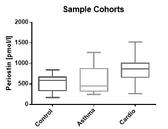
Periostin Values in Asthma and Cardiovascular Disease Panels
In addition to samples from apparently healthy donors, a panel of samples from asthma and cardiovascular disease cohorts were tested.
The results of the experiments are shown below.
| Periostin [pmol/l] | |||||
| Cohort | n | Median | Range | 25% percentile | 75% percentile |
| Control | 18 | 586 | 165 – 840 | 334 | 666 |
| Asthma Cohort* | 10 | 451 | 241 - 1266 | 323 | 869 |
| Cardio Cohort* | 10 | 867 | 260 - 1521 | 663 | 1002 |
*commercially available cohort (Seralab)
Matrix Comparison
Correlation of Serum and Plasma Samples from Apparently Healthy Individuals
14 samples of apparently healthy individuals were prepared as serum and plasma pairs each deriving from one donor. Samples were assayed, and the concentrations of the samples were compared.
|
|
Periostin [pmol/l] | ||||
| Sample ID | Serum | Citrate plasma | EDTA plasma | Heparın plasma | CV (%) |
| #1 | 1297 | 1081 | 1243 | 1195 | 7 |
| #2 | 1303 | 1087 | 1267 | 1345 | 8 |
| #3 | 1710 | 1337 | 1514 | 1512 | 9 |
| #4 | 1946 | 1775 | 1899 | 2251 | 9 |
| #5 | 1539 | 1273 | 1392 | 1108 | 12 |
| #6 | 2279 | 1906 | 2265 | 2316 | 8 |
| #7 | 1901 | 1636 | 1825 | 1712 | 6 |
| #8 | 1879 | 1678 | 1822 | 2028 | 7 |
| #9 | 2295 | 1890 | 2026 | 2102 | 7 |
| #10 | 1572 | 1425 | 1520 | 1760 | 8 |
| #11 | 801 | 669 | 749 | 788 | 7 |
| #12 | 1013 | 883 | 1039 | 806 | 10 |
| #13 | 915 | 796 | 965 | 881 | 7 |
| #14 | 941 | 794 | 992 | 894 | 8 |
| Mean | 8 | ||||
- Periostin as a key molecule defining desmoplastic environment in colorectal cancer. Sueyama T., Kajiwara Y., Mochizuki S., Shimazaki H., Shinto E., Hase K., Ueno H., 2020. Virchows Arch doi: 10.1007/s00428-020-02965-8. Epub ahead of print. PMID: 33215229.
- Chronic Pancreatitis Is Characterized by Elevated Circulating Periostin Levels Related to Intra-Pancreatic Fat Deposition. Ko J., Stuart CE., Modesto AE., Cho J., Bharmal SH., Petrov MS., 2020. J Clin Med Res 12(9):568-578. PMID: 32849945.
- Early effects of androgen deprivation on bone and mineral homeostasis in adult men: a prospective cohort study. Khalil R., Antonio L., Laurent MR., David K., Kim NR., Evenepoel P., Eisenhauer A., Heuser A., Cavalier E., Khosla S., Claessens .F, Vanderschueren D., Decallonne B., 2020. Eur J Endocrinol 183(2):181-189. PMID: 32454455.
- Myostatin and markers of bone metabolism in dermatomyositis. Kerschan-Schindl K., Gruther W., Föger-Samwald U., Bangert C., Kudlacek S., Pietschmann P., 2021. BMC Musculoskelet Disord 5;22(1):150. PMID: 33546660
- Circulating bioactive sclerostin levels in an Austrian population-based cohort. Kerschan-Schindl K., Föger-Samwald U., Gleiss A., Kudlacek S., Wallwitz J., Pietschmann P. , 2021. Wien Klin Wochenschr. Feb 5. doi: 10.1007/s00508-021-01815-0. Epub ahead of print. PMID: 33544208.
- High serum levels of periostin are associated with a poor survival in breast cancer. Rachner, T.D., Göbel, A., Hoffmann, O., Erdmann, K., Kasimir-Bauer, S., Breining, D., Kimmig, R., Hofbauer, L.C., Bittner, A.-K., 2020. Breast Cancer Res Treat.
- Specific effects of anorexia nervosa and obesity on bone mineral density and bone turnover in young women. Maïmoun, L., Garnero, P., Mura, T., Nocca, D., Lefebvre, P., Philibert, P., Seneque, M., Gaspari, L., Vauchot, F., Courtet, P., Sultan, A., Piketty, M.-L., Sultan, C., Renard, E., Guillaume, S., Mariano-Goulart, D., 2019. J Clin Endocrinol Metab.
- Serum periostin levels and severity of fibrous dysplasia of bone.
Guerin Lemaire H., Merle B., Borel O., Gensburger D., Chapurlat R., 2019. Bone 121, 68-71.
PMID:30616028
- Tartrate-resistant acid phosphatase 5b, but not periostin, is useful for assessing Paget’s disease of bone.
Guañabens, N., Filella, X., Florez, H., Ruiz-Gaspá, S., Conesa, A., Peris, P., Monegal, A., Torres, F., 2019. Bone 124, 132–136.
- Characterization of a sandwich ELISA for the quantification of all human periostin isoforms.
Gadermaier, E., Tesarz, M., Suciu, A.A.-M., Wallwitz, J., Berg, G., Himmler, G., 2018. J. Clin. Lab. Anal. 32, e22252.
PMID:28493527
- High circulating levels of Periostin are associated with a poor survival in primary, non-metastatic breast cancer patients.
Hoffmann O, Goebel A, Bittner A-K, Rauner M, Hofbauer LC, Kimmig R, Kasimir-Bauer S, Rachner TD. Proceedings of the 2018 San Antonio Breast Cancer Symposium, 2018. AACR; Cancer Res, 79(4 Suppl): Abstract P6-09-08.
- Effect of Teri paratide Treatment on Circulating Periostin and Its Relationship to Regulators of Bone Formation and BMD in Postmenopausal Women With Osteoporosis.
Gossiel, F., Scott, J.R., Paggiosi, M.A., Naylor, K.E., McCloskey, E.V., Peel, N.F.A., Walsh, J.S., Eastell, R., 2018. The Journal of Clinical Endocrinology & Metabolism 103, 1302–1309.
- Periostin and sclerostin levels in individuals with spinal cord injury and their relationship with bone mass, bone turnover, fracture and osteoporosis status.
Maïmoun, L., Bouallègue, F.B., Gelis, A., Aouinti, S., Mura, T., Philibert, P., Souberbielle, J.-C., Piketty, M., Garnero, P., Mariano-Goulart, D., Fattal, C., 2019. Bone.
PMID:31351195
- Association between non-alcoholic fatty liver disease and bone turnover biomarkers in post-menopausal women with type 2 diabetes.
Mantovani, A., Sani, E., Fassio, A., Colecchia, A., Viapiana, O., Gatti, D., Idolazzi, L., Rossini, M., Salvagno, G., Lippi, G., Zoppini, G., Byrne, C.D., Bonora, E., Targher, G., 2018. Diabetes Metab.
PMID:30315891
- The Utility of Biomarkers in Osteoporosis Management.
Garnero, P., 2017. Mol Diagn Ther 21, 401–418.
- Effect of age and gender on serum periostin: Relationship to cortical measures, bone turnover and hormones.
Walsh, J.S., Gossiel, F., Scott, J.R., Paggiosi, M.A., Eastell, R., 2017. Bone 99, 8–13.
PMID:28323143
 Download biomedica product list


